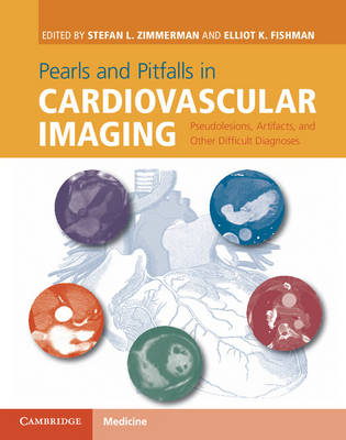
Pearls and Pitfalls in Cardiovascular Imaging
Seiten
2015
Cambridge University Press (Verlag)
978-1-107-02372-7 (ISBN)
Cambridge University Press (Verlag)
978-1-107-02372-7 (ISBN)
- Titel ist leider vergriffen;
keine Neuauflage - Artikel merken
100 real-life cases presented in a succinct and structured format to help cardiovascular imagers avoid common causes of patient misdiagnosis.
Cardiovascular imagers are faced with the challenge of interpreting cases that include artifacts, unusual findings, or anatomic variants on an almost daily basis. These studies can result in confusion and may lead to misdiagnosis even for the most experienced imager.
This book provides an approachable reference for practising cardiovascular imagers to aid with both commonly and uncommonly encountered entities that can result in inappropriate patient management.
Through the focused use of case examples, this book reviews 100 conditions that can be seen in clinical practice, including pseudotumors, artifacts, anatomic variants, mimics, and unusual diagnoses. Each highly illustrated case follows a standard format, allowing readers to learn from real-life examples and provides an accessible and rapid source of reference for the improved interpretation of cardiovascular imaging and enhanced patient care.
This text will be invaluable to radiologists, cardiologists, and trainees.
Stefan L. Zimmerman is Assistant Professor of Radiology and Radiological Sciences, John Hopkins University School of Medicine, Baltimore, MD, USA.
Elliot K. Fishman is Professor of Radiology and Radiological Science, Johns Hopkins University School of Medicine, Baltimore, MD, USA.
Part I. Cardiac Pseudotumors, Tumors, and Other Challenging Diagnoses
Part II. Cardiac Aneurysms and Diverticula
Part III. Cardiovascular Anatomic Variants and Congenital Lesions
Part IV. Coronary Arteries
Part V. Pulmonary Arteries
Part VI. Cardiovascular MRI Artifacts
Part VII. Acute Aorta and Aortic Aneurysms
Part VIII. Post-operative Aorta
Part IX. Mesenteric Vascular
Part X. Peripheral Vascular
Part XI. Veins.
| Erscheint lt. Verlag | 21.5.2015 |
|---|---|
| Zusatzinfo | 615 b/w illus. 39 colour illus. 7 tables |
| Verlagsort | Cambridge |
| Sprache | englisch |
| Maße | 219 x 276 mm |
| Gewicht | 1410 g |
| Einbandart | gebunden |
| Themenwelt | Medizinische Fachgebiete ► Innere Medizin ► Kardiologie / Angiologie |
| Medizin / Pharmazie ► Medizinische Fachgebiete ► Radiologie / Bildgebende Verfahren | |
| Schlagworte | cardiovascular imaging • kardiovaskulär • kardiovaskuläre Magnetresonanztomographie |
| ISBN-10 | 1-107-02372-6 / 1107023726 |
| ISBN-13 | 978-1-107-02372-7 / 9781107023727 |
| Zustand | Neuware |
| Haben Sie eine Frage zum Produkt? |
Mehr entdecken
aus dem Bereich
aus dem Bereich
in Fällen, Fragen und Antworten
Buch | Softcover (2024)
Urban & Fischer in Elsevier (Verlag)
CHF 124,60
Diagnostik und interventionelle Therapie | 2 Bände
Buch (2024)
Deutscher Ärzteverlag
CHF 489,95
Buch | Softcover (2023)
Urban & Fischer in Elsevier (Verlag)
CHF 61,60


