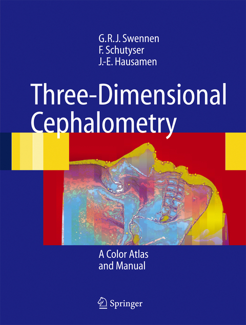
Three-Dimensional Cephalometry
Springer Berlin (Verlag)
978-3-642-06484-5 (ISBN)
Radiographic cephalometry has been one of the most With Three-Dimensional Cephalometry A Color important diagnostic tools in orthodontics, since its Atlas and Manual by the authors Swennen,Schutyser introduction in the early 1930s by Broadbent in the and Hausamen you have an exciting book in your United States and Hofrath in Germany.Generations of hands. It shows you how the head can be analysed in orthodontists have relied on the interpretation of these three dimensions with the aid of 3D-cephalometry. images for their diagnosis and treatment planning as Of course,at the moment the technique is not available well as for the long-term follow-up of growth and in every orthodontic of?ce around the corner. H- treatment results.Also in the planning for surgical ever, especially for the planning of more complex orthodontic corrections of jaw discrepancies, lateral cases where combined surgical orthodontic tre- and antero-posterior cephalograms have been valu- ment is indicated,it is my sincere conviction that wi- able tools.For these purposes numerous cephalomet- in 10 years time 3D cephalometry will have changed ric analyses are available.However,a major drawback our way of thinking about planning and clinical of the existing technique is that it renders only a two- handling of these patients. dimensional representation of a three-dimensional structure.
Prof. em. Dr. Dr. Jarg-Erich Hausamen, ehem. Klinik für Mund,- Kiefer- und Gesichtschirurgie MHH (ehem. Leiter der Akademie für MKG).
From 3-D Volumetric Computer Tomography to 3-D Cephalometry.- Basic Craniofacial Anatomical Outlines.- 3-D Cephalometric Reference System.- 3-D Cephalometric Hard Tissue Landmarks.- 3-D Cephalometric Soft Tissue Landmarks.- 3-D Cephalometric Planes.- 3-D Cephalometric Analysis.- 3-D Cephalometry and Craniofacial Growth.- Clinical Applications.- Future Perspectives of 3-D Cephalometry.
lt;p>Aus den Rezensionen:
"... Der Übergang von der konventionellen Röntgendiagnostik ... in die virtuelle Realität der dreidimensionalen Datensätze war bisher jedoch immer problematisch ... Das vorliegende Buch ... schließt diese Lücke ... Es ist das Verdienst der Herausgeber dieses Buches, in der reich bebilderten Darstellung eine Möglichkeit aufzuzeigen, konventionelle zephalometrische Landmarken in reproduzierbarer Weise auf die dreidimensionale Anatomie des Gesichtsschädels und seiner Weichteile zu übertragen und dort sichtbar zu machen. ... Das Buch hat daher einen hohen praktischen Wert ... Aufgrund seines umfassenden Literaturverzeichnisses ist es ... lohnenswert ..." (Prof. Dr. Dr. H.Schliephake, in: MKG - Mund-, Kiefer- und Gesichtschirurgie, 2006, Vol. 10, Issue 2, S. 126)
| Erscheint lt. Verlag | 12.2.2010 |
|---|---|
| Zusatzinfo | XXII, 366 p. 713 illus., 600 illus. in color. |
| Verlagsort | Berlin |
| Sprache | englisch |
| Maße | 210 x 277 mm |
| Gewicht | 935 g |
| Themenwelt | Medizinische Fachgebiete ► Chirurgie ► Ästhetische und Plastische Chirurgie |
| Medizin / Pharmazie ► Zahnmedizin ► Chirurgie | |
| Schlagworte | 3-D Cephalometry • Assessment • clinical application • Computer • Computer Tomography • Cranofacial • growth • Head • maxillofacial • Orthodontics • Research • Soft tissue • tissue • Tomography |
| ISBN-10 | 3-642-06484-1 / 3642064841 |
| ISBN-13 | 978-3-642-06484-5 / 9783642064845 |
| Zustand | Neuware |
| Haben Sie eine Frage zum Produkt? |
aus dem Bereich


