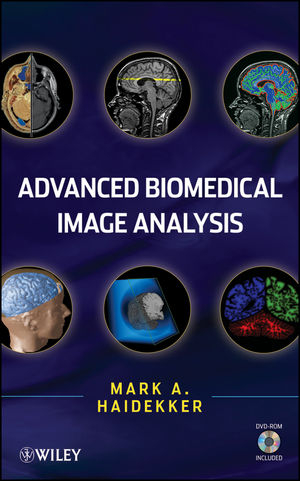
Advanced Biomedical Image Analysis
John Wiley & Sons Inc (Verlag)
978-0-470-62458-6 (ISBN)
- Lieferbar (Termin unbekannt)
- Versandkostenfrei
- Auch auf Rechnung
- Artikel merken
A comprehensive reference of cutting-edge advanced techniques for quantitative image processing and analysis Medical diagnostics and intervention, and biomedical research rely progressively on imaging techniques, namely, the ability to capture, store, analyze, and display images at the organ, tissue, cellular, and molecular level. These tasks are supported by increasingly powerful computer methods to process and analyze images. This text serves as an authoritative resource and self-study guide explaining sophisticated techniques of quantitative image analysis, with a focus on biomedical applications. It offers both theory and practical examples for immediate application of the topics as well as for in-depth study.
Advanced Biomedical Image Analysis presents methods in the four major areas of image processing: image enhancement and restoration, image segmentation, image quantification and classification, and image visualization. In each instance, the theory, mathematical foundation, and basic description of an image processing operator is provided, as well as a discussion of performance features, advantages, and limitations. Key algorithms are provided in pseudo-code to help with implementation, and biomedical examples are included in each chapter.
Image registration, storage, transport, and compression are also covered, and there is a review of image analysis and visualization software.
Members of the academic community involved in image-related research as well as members of the professional R&D sector will rely on this volume.
It is also well suited as a textbook for graduate-level image processing classes in the computer science and engineering fields.
MARK A. HAIDEKKER, PHD, is an associate professor in the Faculty of Engineering at the University of Georgia. Dr. Haidekker develops image analysis methods based on modern imaging modalities, including computed tomography (CT), magnetic resonance imaging (MRI), and optical imaging. Specific areas of his research include the relationship between images of organs and their biomechanical properties, the development and improvement of laser-based imaging modalities, and the development of methods to image microviscosity and micro-flow patterns with mechanosensitive fluorescent molecular rotors.
Preface ix
1 Image Analysis: A Perspective 1
1.1 Main Biomedical Imaging Modalities 3
1.2 Biomedical Image Analysis 7
1.3 Current Trends in Biomedical Imaging 12
1.4 About This Book 15
References 17
2 Survey of Fundamental Image Processing Operators 23
2.1 Statistical Image Description 24
2.2 Brightness and Contrast Manipulation 28
2.3 Image Enhancement and Restoration 29
2.4 Intensity-Based Segmentation (Thresholding) 42
2.5 Multidimensional Thresholding 50
2.6 Image Calculations 54
2.7 Binary Image Processing 58
2.8 Biomedical Examples 63
References 68
3 Image Processing in the Frequency Domain 70
3.1 The Fourier Transform 71
3.2 Fourier-Based Filtering 82
3.3 Other Integral Transforms: The Discrete Cosine Transform and the Hartley Transform 91
3.4 Biomedical Examples 94
References 100
4 The Wavelet Transform and Wavelet-Based Filtering 103
4.1 One-Dimensional Discrete Wavelet Transform 106
4.2 Two-Dimensional Discrete Wavelet Transform 112
4.3 Wavelet-Based Filtering 116
4.4 Comparison of Frequency-Domain Analysis to Wavelet Analysis 128
4.5 Biomedical Examples 130
References 135
5 Adaptive Filtering 138
5.1 Adaptive Noise Reduction 139
5.2 Adaptive Filters in the Frequency Domain: Adaptive Wiener Filters 155
5.3 Segmentation with Local Adaptive Thresholds and Related Methods 157
5.4 Biomedical Examples 164
References 170
6 Deformable Models and Active Contours 173
6.1 Two-Dimensional Active Contours (Snakes) 180
6.2 Three-Dimensional Active Contours 193
6.3 Live-Wire Techniques 197
6.4 Biomedical Examples 205
References 209
7 The Hough Transform 211
7.1 Detecting Lines and Edges with the Hough Transform 213
7.2 Detection of Circles and Ellipses with the Hough Transform 219
7.3 Generalized Hough Transform 223
7.4 Randomized Hough Transform 226
7.5 Biomedical Examples 231
References 234
8 Texture Analysis 236
8.1 Statistical Texture Classification 238
8.2 Texture Classification with Local Neighborhood Methods 242
8.3 Frequency-Domain Methods for Texture Classification 254
8.4 Run Lengths 257
8.5 Other Classification Methods 263
8.6 Biomedical Examples 265
References 273
9 Shape Analysis 276
9.1 Cluster Labeling 278
9.2 Spatial-Domain Shape Metrics 279
9.3 Statistical Moment Invariants 285
9.4 Chain Codes 287
9.5 Fourier Descriptors 291
9.6 Topological Analysis 295
9.7 Biomedical Examples 301
References 307
10 Fractal Approaches to Image Analysis 310
10.1 Self-Similarity and the Fractal Dimension 311
10.2 Estimation Techniques for the Fractal Dimension in Binary Images 319
10.3 Estimation Techniques for the Fractal Dimension in Gray-Scale Images 327
10.4 Fractal Dimension in the Frequency Domain 331
10.5 Local Hölder Exponent 337
10.6 Biomedical Examples 340
References 345
11 Image Registration 350
11.1 Linear Spatial Transformations 352
11.2 Nonlinear Transformations 355
11.3 Registration Quality Metrics 360
11.4 Interpolation Methods for Image Registration 371
11.5 Biomedical Examples 379
References 382
12 Image Storage Transport and Compression 386
12.1 Image Archiving DICOM and PACS 389
12.2 Lossless Image Compression 392
12.3 Lossy Image Compression 400
12.4 Biomedical Examples 408
References 411
13 Image Visualization 413
13.1 Gray-Scale Image Visualization 413
13.2 Color Representation of Gray-Scale Images 416
13.3 Contour Lines 422
13.4 Surface Rendering 422
13.5 Volume Visualization 427
13.6 Interactive Three-Dimensional Rendering and Animation 433
13.7 Biomedical Examples 434
References 438
14 Image Analysis and Visualization Software 441
14.1 Image Processing Software: An Overview 443
14.2 ImageJ 447
14.3 Examples of Image Processing Programs 452
14.4 Crystal Image 456
14.5 OpenDX 461
14.6 Wavelet-Related Software 466
14.7 Algorithm Implementation 466
References 473
Appendix A: Image Analysis with Crystal Image 475
Appendix B: Software on DVD 497
Index 499
| Erscheint lt. Verlag | 3.12.2010 |
|---|---|
| Verlagsort | New York |
| Sprache | englisch |
| Maße | 163 x 241 mm |
| Gewicht | 980 g |
| Themenwelt | Informatik ► Grafik / Design ► Digitale Bildverarbeitung |
| Medizin / Pharmazie ► Medizinische Fachgebiete ► Radiologie / Bildgebende Verfahren | |
| Technik ► Elektrotechnik / Energietechnik | |
| ISBN-10 | 0-470-62458-2 / 0470624582 |
| ISBN-13 | 978-0-470-62458-6 / 9780470624586 |
| Zustand | Neuware |
| Haben Sie eine Frage zum Produkt? |
aus dem Bereich


